

Normal Aortic Valve (II)
 Aortic Root and Aortic Valve This image of the aortic root and the aortic valve shows greater detail of these structures.
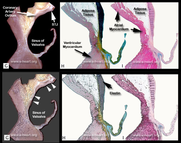
G. Gross photograph of a parasagittal section through the aortic root showing the aorta as it approaches the junction of the left anterior atrial wall with the ventricular septum. The fibrous annulus is thin at this point. The aorta becomes thin as it approaches the sinotubular junction and merges into the aortic leaflet.
H. Histologic section of the specimen shown in G. Note the prominent elastic lamellae (black) in the aortic wall. These lamellae end abruptly at the point where the aortic root merges with the fibrous annulus (yellow). The base of the leaflet has abundant proteoglycan material (green) and some elastic fibers within the spongiosa. (Movat pentachrome, 3X)
I. Histologic section of the same specimen shown in H. Note the small strand of muscle fibers that are located beneath the fibrous annulus, course caudally in the subendocardium, and represent the left posterior fascicle of the bundle of His. (H&E, 3X)