

Cardiac histology (II)
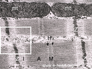 The Z bands are clearly visible. Adjacent to the Z bands there are lighter areas (I bands, I = stands for Isotropic, when the striated muscle is analyzed under polarized light) which are areas where the thin filaments attach to the Z band. Between the I bands there is a large darker gray area called the A band (A stands for anisotropic). The A band is the place where the thin and thick filaments overlap and interact during contraction of the myocyte. The M band is the conspicuous middle line of the A band. To each side of the M band there is an area of lighter material (less electron dense) which is the H band (arrows). In the 3D Visualization area you may see further detail of this and also the molecules that form each one of these structures in the myocyte.
The white rectangle shows and area between to myofibrils, at the level of the Z bands. In these region there are cytoskeletal (intermediate 10 nm) filaments made of desmin in the mature cardiac myocytes.
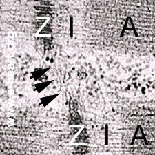 A higher magnification of the cytoskeletal intermediate filaments is shown here. The arrows point to the 10 nm-thick intemediate filaments that join adjacent Z disks. These intermediate filaments are made of Desmin.
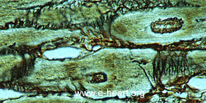 This micrograph shows that in a similar fashion to the the function of the cytoskeletal filaments maintaining the sarcomeres in register, the extracellular matrix of the heart plays a role in maintaining the orderly array of the cardiac myocytes. Collagen bundles form bridges between the basal laminae of adjacent myocytes (struts and cables) and also between myocytes an capillaries. This reticulum stain shows such bundles of collagen (black lines) forming bridges between adjacent myocytes.
The structure of the myocytes shown so far, applies to all working myocytes. However there are specialized myocytes in the myocardium including the conduction system myocytes in the sinoatrial node (SA Node), the atrioventricular node (AV Node) the His Bundle and its branches and the Purkinje system.
Also the atrial myocytes have specific features such as atrial specific granules where atrial natriuretic peptide is stored.