

Mitral Valve (II)
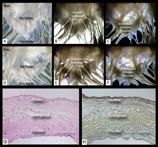 The atrial aspect of the posterolateral leaflet of a normal mitral valve is shown in panels A to C and the ventricular aspect of the same valve is shown in panels D to F. Transillumination (B and E) and examination under polarized light (C and F) show areas of different density which correspond to the insertion sites of different types of chordae tendineae. The leaflet can be divided into three zones: a rough zone where first and second order chordae insert, a clear zone, and a basal zone where third order chordae insert. The collagen bundles follow the general direction of the chordae with which they are continuous. Also note the strong birefringence of the chordae.
F and G show that upon light microscopic examination there are three discrete layers in the atrioventricular valves. These layers vary slightly according to the region (whether basal or distal) of the leaflet. The fibrosa is the most consistent layer being found from the base to the free edge. It is formed by collagenous bundles (yellow). The spongiosa contains loosely arranged connective tissue, proteoglycans and small amount of elastic fibers. It is most prominent in the free edge of the valve. At the basal portion, it contains extension of left atrial myocardium. A few adipose tissue cells may be seen within this layer occasionally. The atrialis or auricularis covers the atrial aspect of the spongiosa in the basal and mid portion of the valve. It is also rich in proteoglycans and contains elastic fibers and occasional smooth muscle cells. With age, fibrosis (sclerosis) becomes the predominant change at the base and myxoid degeneration at the free edge. However excessive accumulation of glycosaminoglycans (GAG's also known as mucopolysaccharides) in the spongiosa is commonly seen in mitral valve prolapse. The chordae tendienae insert into the fibrosa layer of the valve. Normal atrioventricular valves are avascular, and the presence of vessels within the valve is a pathologic change. (H&E and Movat pentachrome, 400X).
For comparison with the tricuspid valve you may see the common features of the atrioventricular valves here.
Back to cardiac structure