

Cardiac Amyloidosis - VI
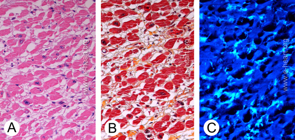
Isolated atrial amyloid. Autopsy exam showed slight rigidity of the left atrial wall. A. The cardiac myocytes show hypertrophy but no distinct eosinophilic infiltrate in the interstitial space. (H&E X 400) B. Interstitial (perimysial) fibrous tissue (collagen) is highlighted in yellow and a very faint green discoloration is noted in some the fibrous strands of interstitial fibrous tissue. (Movat stain X 400) C. Examination of the Thioflavin-S stain under UV light microscopy shows fine strands of amyloid (light blue strands) corresponding to the areas of green discoloration in the Movat stain. (Thioflavin-S X400)
The morphologic patterns of amyloid seen in the ventricles in clinically symptomatic patients are intersitial , arteriolar , nodular , venular and can also involve the cardiac adipose tissue.