

Normal Pericardial Anatomy - Phrenic Nerves - V
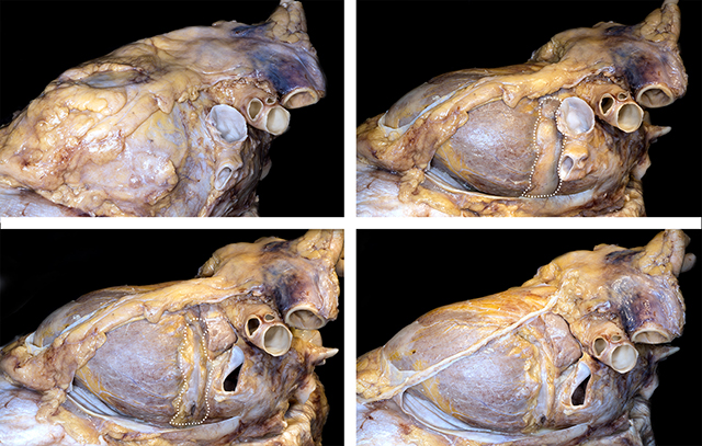
These images show the left phrenic nerve as it courses over the parietal pericardium (top left). Partial removal of the anterior and lateral portions of the parietal pericardium show the epicardial surface of the heart. The dotted line highlights the atrioventricular groove with abundant adipose tissue. Beneath the adipose tissue the are the great cardiac vein and the coronary sinus (top right). The lower left image show the atrioventricular grove adipose tissue and thin layer of epicardium and lateral portion of vein wall removed to demonstrate the lumen of the great cardiac vein and coronary sinus. The lower right images shows further dissection of the adipose tissue that covers the phrenic nerve.
The close relationship between the parietal (fibrous) pericardium and visceral pericardium as well as the pericardial space are shown here.
Back to Pericardial disease
Back to Home Page