

Pericarditis - Organizing - II
 Acute fibrinous pericarditis. A and B. Fibrinous exudates with abundant inflammatory cells are shown. The fibrosa layer shows dilated vessels. The inset shows a close up of the inflammatory infiltrates intermingled with the fibrinous strands and reactive mesothelial cells in the serosa layer of the parietal pericardium.
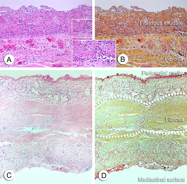
C and D. Parietal pericardium with abundant inflammatory infiltrates and early deposition of extracellular matrix (yellow green color in D on top and below the fibrosa). This is an early stage of organization of the exudate. The newly formed connective tissue eventually is invaded by capillaries that form extensive vascular network. The fibrosa layer of the parietal pericardium is delineated by the dotted lines. Note that in this example the inflammatory process is involving both the pericardial serosa (mesothelial) layer as well as the mediastinal pleural serosa layer. If a pericardiectomy is performed, the inflammation present in the mediastinal pleura may still present clinically as residual pericardial referred pain.
(A and C. H&E stain. B and D. Movat pentachrome stain).
Further stages of organization show more mature fibrious tissue with less inflammation.
Back to Pericardial disease