Arrhythmogenic Right Ventricular Dysplasia / Cardiomyopathy - ARVD / C

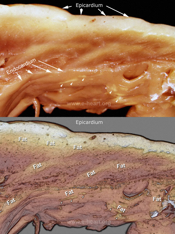
Section of the right ventricular free wall is shown. The epicardial surface is marked (arrow). The cut surface of the right ventricular wall shows fatty infiltration seen as yellow areas alternating with the normal red-brown color of the myocardium. The endocardium is marked by arrows. There are multiple yellow streaks (arrowheads) clearly noticeable on the endocardial surface due to adipose tissue deposition. The lower image is an illustration highlighting the areas of adipose tissue witing the myocardium of this section through the wall of the right ventricle. Pathologic examination of the heart with suspected ARVD should be very thorough, since there is a wide variation of the normal amount of adipose tissue infiltration in humans.
(Tansey DK, Aly Z, Sheppard MN : Fat in the right ventricle of the normal heart.
Histopathology. 2005 ;46: 98-104.)
The diagnosis of ARVD in endomyoardial biopsies can be suspected if abundant adipose tissue is present. Concomitant fibrosis and the presence of inflammtory cells can be seen. But the definitive diagnosis requires clinical and imaging correlation.
( Castillo E, Tandri H, Rodriguez ER, Nasir K, Rutberg J, Calkins H, Lima JA, Bluemke DA: Arrhythmogenic Right Ventricular Dysplasia: Ex Vivo and in Vitro Fat Detection with Black-Blood MR Imaging. Radiology 2004: 232; 38-48
Tandri H, Saranathan M, Rodriguez ER, Martinez C, Bomma C, Nasir K, Rosen B, Lima JA, Calkins H, Bluemke DA: Noninvasive detection of myocardial fibrosis in arrhythmogenic right ventricular cardiomyopathy using delayed-enhancement magnetic resonance imaging. J Am Coll Cardiol 2005: 45; 98-103
Fritz J, Tandri H, Rodriguez ER, Calkins H Bluemke DA: Evaluation and course of an unusual case of arrhythmogenic right ventricular dysplasia. Int J Cardiovasc Imaging. 2005; 22: 269-73.
)

