Cardiac Amyloidosis II

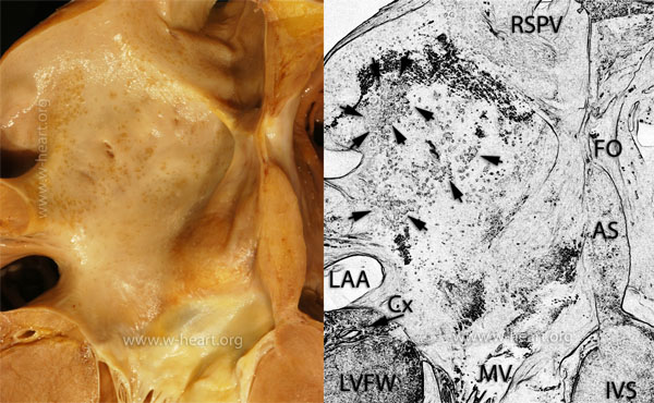
This gross image shows the endocardial surface of the anterior wall of the left atrium. The interatrial septum is visible on the right side of the image (AS). The Fossa Ovalis (FS) is rather distinct. The cephalad portion of the anterior leaflet of the mitral valve (MV) is present at the bottom of the image. The interventricular septum (IVS) and the left ventricular free wall (LVFW) are also labeled. The circumflex coronary artery (Cx) is noted in the atrioventricular groove (arrow). The opening of the left atrial appendage (LAA) (lower left) and the right superior pulmonary vein (RSPV) are labeled. Multiple arrows point at the prominent glassy (ochre-yellow) discoloration areas in the atrial endocardium which correspond to nodules of amyloid deposits. The amyloid deposits can appear as small grains (sandpaper like) in the right atrium or nodules and plaques in the left atrium and valves.

