Hypertrophic Cardiomyopathy - HCM - LAMP2
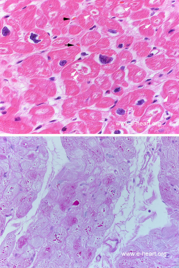
 The upper panel shows a hematoxylin & eosin stain in which there is marked myocyte hypertrophy, and bizarre-shaped myocyte nuclei are present. The arrows point at eosinophilic inclusion present in the myocytes. The intra-sarcoplasmic inclusions are distinctly different from red blood cells (black arrows).
The lower panel is a positive PAS stain showing the strong reactivity of the carbohydrate moieties with the Schiff reagent (intense pink deposits and granules). The ultrastructure of these inclussions which have been complared to phagolysosomes is shown on transmission electron microscopy.

