Histology of the Pericardium - II
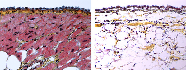
 C. The visceral pericardium (also called epicardium) consists of a mesothelial cell layer and a thin subepithelial layer of collagen with elastic fibers. The mesothelial cells show distinct microvilli. Beneath the visceral pericardium is epicardial adipose tissue. D. In other areas of the visceral pericardium (or epicardium), only a thin layer of fibrous tissue (yellow) separates the mesothelial cells from the myocardium with absence of epicardial fat.

These two microscopic images the serosal (mesothelial cell) component of the visceral pericardium that invests the heart. The image on the left represents an area where the visceral pericardium is in direct contact with the myocardium. Only a thin layer of fibrous tissue (yellow) separates the mesothelial cells (grey blue cytoplasm) from the cardiac myocytes (red sarcoplasm). Note the scant amount of elastic lamellae shown as black straight lines in the middle of the yellow fibrous tissue just underneath the mesothelium. (X200, Movat pentachrome) The image on the right shows the visceral pericardium as it covers an area of the heart with abundant adipose tissue in the epicardium (such as the interventricular and atrioventricular grooves or around the coronary vessels). There is a small amount of fibrous tissue (yellow) as well as scant elastic lamellae (black). The mesothelial cells forming the serosal layer play important role in the production and reabsorption of fluid in the pericardial space. (X200, Movat pentachrome).
The ultrastructural features of the mesothelial cells that form the serosal pericardium are shown here.
Back to Pericardial disease
Back to Home Page

