

Molecular Anatomy of the Heart (I)
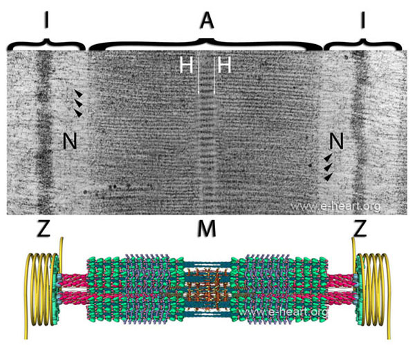
The sarcomere. The upper panel shows a transmission electron micrograph of a human cardiac sarcomere. The boundaries of the sarcomere are the 2 Z-discs (Zwischencheibe) which are dark and electron dense. On each side of the Z-discs, there is an area of electron-lucent material with fine filaments that run perpendicular to the Z-disc. These represent actin filaments which insert into the Z-disc. The area that encompasses a Z-disc and adjacent electron-lucent area of actin filaments from two sarcomeres is designated the I band (Isotropic band). In the middle of this electron-lucent actin rich area, there are some subtle electron-dense streaks that run parallel to the Z-disc and are named the N-disc (Nebenscheibe) . In this area, there are some proteins that interact with titin, actin and α-actinin. The mid portion of the sarcomere is comprised of the A band (Anisotropic band). Within the A band, there are several areas with specific patterns of electron density. There is a middle line or M band (Mittellinie) which bisects the A band. Adjacent to the M band, there are two lighter electron-lucent areas designated the H band (Helle). The M band corresponds to the insertion site of the tails of the thick filaments into a matrix of proteins that keep them organized and in register. Within the M band, there are subtle electron-dense core of proteins which correspond to myomesin and other proteins that serve to anchor the thick filaments. The larger dark areas that flank the M and H bands are the actual rows of thick filaments. In the middle third of these areas, there are some vertical striations which correspond to electron-dense myosin binding protein C. The lower portion of the figure shows a simplified illustration of the sarcomere. The M band shows the tails of the thick filaments interacting with a light brown lattice of myomesin molecules. The thick filaments extend away from the M band with the heads (green) protruding away from the axis of the filament. Myosin binding protein C is visible as small blue subunits that cover about one third of the length of the thick filament. The thin filaments (red) are made of several proteins (see The Thick and Thin Filaments). The yellow coils shown at the level of the Z-disc represent intermediate filaments of desmin which connect adjacent myofibrils to each other and to the sarcolemma. An animation of the sarcomere contraction is shown here and here http://www.youtube.com/watch?v=-pg09F5V63U.