

Right Ventricle (II)
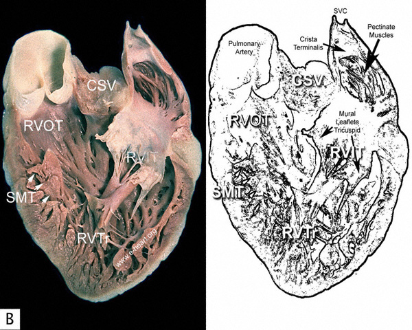 B . This is a view of the inner aspect of the right atrial and ventricular free walls,
which have been dissected from the specimen shown in A to demonstrate
the inflow apical and outflow regions. The tricuspid valve is separated
from the pulmonic valve by a muscular fold, which forms the roof of the
crista supraventricularis (CSV). (SMT= Septomarginal Trabecula) (RVTr = Right ventricle, trabecular portion) (RVIT = Rigth Ventricular Inflow
Tract) (RVOT = Right Ventricular Outflow Tract)
B . This is a view of the inner aspect of the right atrial and ventricular free walls,
which have been dissected from the specimen shown in A to demonstrate
the inflow apical and outflow regions. The tricuspid valve is separated
from the pulmonic valve by a muscular fold, which forms the roof of the
crista supraventricularis (CSV). (SMT= Septomarginal Trabecula) (RVTr = Right ventricle, trabecular portion) (RVIT = Rigth Ventricular Inflow
Tract) (RVOT = Right Ventricular Outflow Tract)
Note the coarse trabeculations (trabeculae carneae) typical of the right ventricle.
The following page shows an anterior view of the crista supraventricularis