

Right Ventricle (I)
 Right ventricle. A. View of the septal components of the right atrium and right ventricle. The free walls of the right atrium and right ventricle have been removed to fully expose the septal and outflow portions of the right ventricle. The ventricle is divided into three segments: 1) the inflow tract or inlet (RVIT); 2) the apical or trabecular portion (RVT), and 3) the outflow tract or outlet (RVOT). The inflow tract is posteroinferior and includes the tricuspid valve apparatus. The apical portion is coarsely trabeculated. The outflow tract, also called the conus arteriosus or infundibulum, is located anterosuperiorly and is a muscular structure that supports the pulmonic valve.
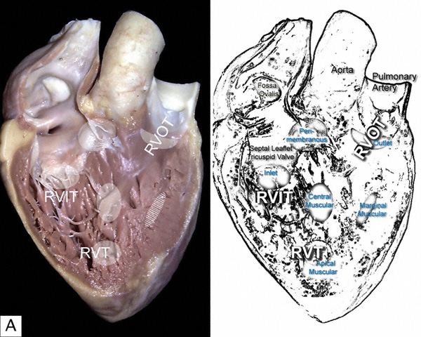
The areas shaded in white represent common sites for interventricular septal defects, which are designated according to their topography. From the inflow to the outflow tract they are: inlet defects; perimembranous; central muscular defects; apical muscular defects; marginal muscular defects; and outlet defects.
The following image illustrates the inner aspect of the lateral walls of the right atrium and and right ventrice.