

Myocardial infarct - Ventricular Aneurysm
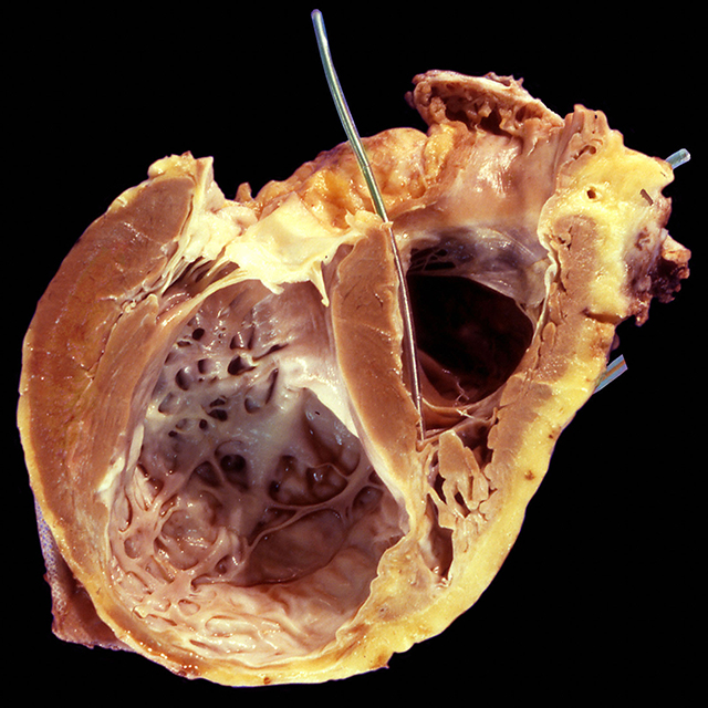
This image shows an explanted heart seen the dorsal-inferior plane. THe left ventricle shows compensatory hypertrophy of the basal portion of the laeral wall and interventricular septum. An apical aneurysm is present. The aneurysm shows a white thickened endocardium (endocardial fibroelastosis) and also a thrombus entangled in the thin trabeculae. The ventricular wall is only a few milimeters thick int his area. A pacemaker lead is present in the right venttricle and it is anchored near the apex of this chamber.
Back to Myocardial Infarct
Back to Home Page