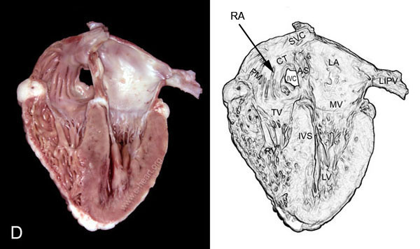
Four Chamber View of the Heart

This is a four chamber view of the heart. The right atrium (RA) shows multiple muscular trabeculation (pectinate muscles - PM). The pectinate muscles form a structural "vault" to keep the shape of the thin-walled right atrium expanded, and they coalesce into the crista terminalis (CT). In contrast to the right atrium, the left atrium has a rather smooth endocardial surface with no trabecular muscle bundles. Note the contrast of the endocardium of the left atrium (white) to the endocardium of the right atrium. The posterior leaflet of the tricuspid valve (TV) is shown in the image. The right ventricle (RV) shows abundant coarse trabeculations throughout its surface.
The Mitral Valve (MV) shows that its chordae tendineae are anchored to the posterolateral and the anteromedial papillary muscles. Compare the thickness of the right ventricular free wall to the thickness of the left ventricular free wall.
The superior and inverior venae cavae (SVC and IVC) open into the right atrium. The left inferior pulmoary vein (LIPV) is shown opening into the left atrium.

