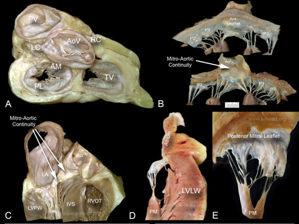
Mitral Valve (I)
 A. Gross specimen with a cephalad view of the base of the heart after removing the atria. The epicardial fat is still present and the coronary arteries (LC and RC) are visible. The aortic valve occupies a central position at the base of the heart and anchors the other three valves. The aortic root is at the center of the fibrous skeleton of the heart which provides attachment for the leaflets of the aortic, mitral and tricuspid valves as well as for the atrial and ventricular myocardium. Note that the pulmonic valve has an orientation opposite to that of the aortic valve, due to a criss-cross arrangement of the two outflow tracts.
B. The atrial and ventricular surfaces of the mitral valve are shown. The mitral valve consists of an anterior (aortic, septal, anteromedial) and a posterior (mural, ventricular, posterolateral) leaflets. The anterior leaflet is roughly triangular and occupies one-third of the mitral valve circumference. The posterior leaflet is longer with shallow indentations creating scallops that are often referred to as P1, P2 and P3. In the lower image in B, the anterior mitral leaflet is seen in continuity with the aortic valve. Commissures have corresponding papillary muscles that anchor the fan-shaped chordae. In the mitral valve, the chordae tendineae are anchored by the anterolateral and posteromedial papillary muscles. Different pathologic conditions affect these subcomponents of the valve. In mitral valve prolapse (also called myxomatous degeneration) there is weakness of the leaflet and chordal connective tissue. Inflammatory diseases such as rheumatic fever can produce injury to the leaflets, the commissures and the chordae tendineae.
C. In contrast to the right ventricle, where the tricuspid and pulmonic valves are separated by a muscular band, the left ventricle is characterized by fibrous( mitro-aortic) continuity between the mitral and the aortic valves. The anterior mitral leaflet, being continuous with the left and posterior (non coronary) aortic cusps, divides the left ventricle into inflow and outflow portions. Age-related changes affecting the valves that are evident in this specimen include thickening or sclerosis of the valve leaflets and calcification that begins in the mitral annulus. Minor degrees of mitral calcification are usually of no clinical significance. When severe, it forms nodular masses of calcium that extend deeply into the myocardium and produce calcific spurs on the ventricular aspect of the leaflets. The aortic root is mildly dilated and shows the sinotubular junction, a ridge formed at the border between the sinuses and the tubular segment of the ascending aorta.
D. The atrial surface of atrioventricular valve leaflets is smooth with variable degrees of ballooning that ensure tight apposition during systole. The ventricular surface is marked by chordal attachment to different zones of the leaflet. Chordae arising from the heads of papillary muscles can insert on the free edge (first order) or on the ventricular mid zone (second order) of the leaflets. Chordae that originate from trabeculae carneae and insert on the basal zone of the leaflets are called chordae of the third order.
E. For comparison to the mitral valve, the ventricular aspect of a tricuspid valve is shown to demonstrate chordal insertion to the free margin and mid portion of the leaflet. The basal portion is clear and receives little or no chordae. The chordae in the tricuspid valve are thinner that in the mitral valve.
A detailed view of the mitral leaflet structure and histology is shown here.

