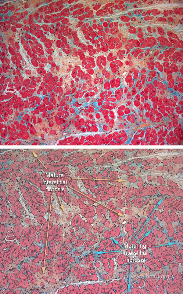
Myocardial interstitial fibrosis I
 Interstitial fibrosis is illustrated in this Movat pentachrome stain. The cardiac myocytes (stained in red) are mostly sectioned in a transverse plane. This highlights the extracellular matrix. The mature collagen bundles are stained in yellow / ochre, and the more immature fibrous tissue shows some proteoglycan material (blue / green). This extracellular matrix encases the myocytes and restricts their contractility.
Interstitial fibrosis can be associated to concomitant endocardial fibroelastosis in some cases

