Normal Pericardial Anatomy - IV

Parietal and visceral pericardium.
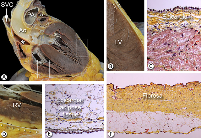
A. Coronal section of the heart shows the ventricles, ascending aorta (Ao), and partial views of the right and left atrial appendages, the superior vena cava (SVC), the aortic valve and the pulmonary artery trunk (PA).
B. The inset of the left lateral ventricular wall is magnified. The visceral and parietal pericardium are in close apposition and the space between these two layers is virtual. The arrowheads show an area of folding of the parietal pericardium where it separates from the visceral pericardium. Note the lack of subepicardial fat in the lateral left ventricle.
C. Light microscopic exam shows a thin layer of fibrous tissue (yellow) overlying the cardiac muscle (red). The “hobnail” cells lying over the thin fibrous sheet are the mesothelial cells which form the visceral pericardium. Note the close proximity of the myocardial capillaries to the mesothelium of the visceral layer. This rich network of vessels can provide fast transfer of fluid material in and out of the pericardial space.
D. A close up of the inset on the right ventricle in 3A is shown. There is a distinct space between the parietal pericardium (arrowheads) and the epicardium covering the adipose tissue that overlies the right ventricular myocardium (RV).
E. Light microscopy of the mesothelial lining over the adipose tissue of the right ventricle (visceral pericardium). Short elastic fibers (black) are present in the subepicardium.
F. This micrograph of full thickness pericardial sac shows the fibrosa layer of the parietal pericardium. Note the sparse vascularization of the fibrosa. The mesothelial cells of the parietal pericardium are directly attached to the fibrosa in the upper part of the photo. The mediastinal aspect (lower part) of the pericardium shows adipose tissue which, in turn, is also covered by mesothelial cells forming the serosa of the mediastinal pleura.
Ao = aorta. LV = left ventricle. PA = pulmonary artery. RV = right ventricle. SVC = superior vena cava. * = inferior aortic recess. ** = Left pulmonic recess. A close up of the recesses is shown here.
 A and C. The parietal pericardium is well delineated by the epipericardial fat on the anterior diaphragmatic surface and epicardial fat over the right ventricle.
B and D. The left ventricle contains little epicardial fat resulting in poor visualization of the pericardium in this area on imaging.
RV = Right ventricle. LV = Left ventricle. Large asterisks = mediastinal adipose tissue over the fibrous pericardium. Small asterisks = epicardial fat over the RV and LV. Dashed red lines = Pericardial space (virtual in the normal heart).
The transverse and oblique sinuses of the pericardium are illustrated in right lateral and left lateral views here.
Back to Pericardial disease
Back to Home Page

