Normal Pericardial Anatomy - I

The pericardium, a roughly flask-shaped sac that contains the heart, consists of a fibrous and a serosal component. This sac surrounds the heart and the vessels that enter and exit the heart (i.e. Aorta, pulmonary artery, venae cavae, and pulmonary veins). The areas where the pericardial sac attaches to these vessels form sinuses (oblique and transverse) as well as recesses (aortic and pulmonic).
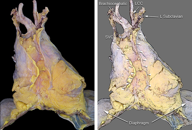
In these images A and B the anterior view of the intact parietal (fibrous) pericardial sac is shown. The attachment of the fibrous sac to the diaphragm is seen at the base. Abundant epipericardial fat is conspicuously present at the pericardial-diaphragm junction. The mediastinal pleura invest the lateral portion of fibrous pericardium. The anterior reflections of the mediastinal pleura are indicated by the white arrowheads. The space between the arrowheads corresponds to the attachment of the pericardium to the posterior surface of the sternum. Superiorly, the left innominate vein is seen merging with the superior vena cava. The arterial branches of the aortic arch are just dorsal to the innominate vein.
C and D. The anterior portion of the pericardial sac (fibrous pericardium) has been removed to show the heart and great vessels in anatomic position. It distinctly shows how the proximal segments of the great arteries are intrapericardial. A that point, there is fusion of the adventitia of the great vessels with the fibrous pericardium.
SVC = Superior vena cava. Ao = Aorta. PA = Pulmonary artery. LAA = Left atrial appendage. RAA = Right atrial appendage. RV = Right ventricle. LV = Left ventricle.

This image shows an intact pericardium attached to the diaphragm is shown with the mediastinal pleura covering the lateral surfaces of the pericardial sac. Note the abundant epipericardial adipose tissue in the anterior mediastinum and anterior cardiophrenic angles. The arrowheads flank the borders of the sterno-pericardial ligament.
LCC = Left common carotid artery. SVC = Superior vena cava.
The right lateral and left lateral views of the pericardium are shown here.
Back to Pericardial disease

